Ultrasound-guided sensory-predominant block for anteromedial knee analgesia (saphenous nerve ± nerve to vastus medialis) with relative quadriceps sparing
Surgeries where ACB is commonly used:
- Total knee arthroplasty (TKA)
- Midline skin incision, medial parapatellar arthrotomy, bone cuts and implant cementation, tourniquet may be used, closure with drains variably used
- ACL reconstruction / knee arthroscopy / meniscal surgery
- Arthroscopic portals, graft harvest (hamstring/patellar tendon) may add pain, usually day-case
- Medial knee procedures (e.g. unicompartmental knee arthroplasty, medial tibial plateau fixation)
- Analgesic requirement varies with bony work and tourniquet duration
Definition and aims
- Ultrasound-guided injection of local anaesthetic in the adductor canal to block the saphenous nerve (terminal sensory branch of femoral nerve) ± nerve to vastus medialis and other small articular branches
- Aim: analgesia for anteromedial knee and medial leg/ankle with relative preservation of quadriceps strength compared with femoral nerve block
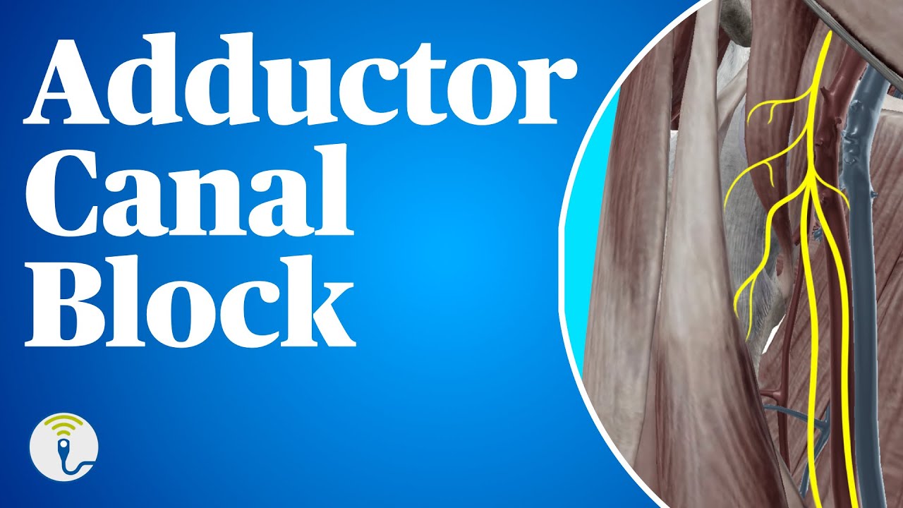
Relevant anatomy
- Adductor canal (subsartorial/Hunter’s canal): aponeurotic tunnel in mid-thigh
- Boundaries: roof—sartorius, lateral—vastus medialis, posterior/medial—adductor longus (proximal) and adductor magnus (distal)
- Contents: femoral artery and vein, saphenous nerve, nerve to vastus medialis (often), subsartorial plexus (variable)
- Saphenous nerve: sensory, accompanies femoral artery in canal then becomes superficial between sartorius and gracilis, supplies medial leg and foot (to first MTP region variably)
- Key concept: ACB does not reliably cover posterior knee (tibial nerve/popliteal plexus) → consider iPACK or other adjuncts for TKA
Indications
- Analgesia for knee surgery: TKA, unicompartmental knee arthroplasty, ACL reconstruction, arthroscopy (selected), medial knee procedures
- Analgesia for medial leg/ankle procedures (saphenous territory) as adjunct (e.g. medial malleolus surgery) when combined with sciatic/popliteal block
Contraindications (as for peripheral nerve blocks)
- Patient refusal, inability to consent/cooperate
- Infection at injection site, systemic sepsis (relative)
- Allergy to local anaesthetic
- Coagulopathy/anticoagulation: follow regional anaesthesia anticoagulation guidance, ACB is a deep peripheral block in proximity to femoral vessels, treat with caution
- Pre-existing neuropathy: relative, document baseline deficits and counsel re: attribution of symptoms
Equipment and preparation
- Ultrasound: high-frequency linear probe (typical adult), consider curvilinear in very large thighs
- Needle: 50–100 mm echogenic regional needle, in-plane approach preferred
- Asepsis: chlorhexidine/alcohol skin prep (allow to dry), sterile probe cover/gel, sterile gloves
- Monitoring and resuscitation: standard monitoring, IV access, intralipid available, LA toxicity plan
Technique (ultrasound-guided, mid-thigh ACB)
- Position: supine, leg slightly externally rotated, expose mid-thigh
- Probe placement: transverse at mid-thigh to identify femoral artery deep to sartorius, vastus medialis lateral, adductors posterior/medial
- Target: perivascular space anterolateral to femoral artery beneath sartorius (within canal), saphenous nerve may be seen as hyperechoic structure
- Needle: in-plane lateral-to-medial (common) or medial-to-lateral, advance to just adjacent to artery within canal, aspirate and inject incremental aliquots observing spread
- Volume: commonly 10–20 mL (institution dependent), higher volumes increase spread (including potential proximal femoral nerve spread) and risk of motor block
- Continuous catheter option: place catheter 3–5 cm beyond needle tip within canal, secure and run low-concentration LA infusion (local protocol)
Local anaesthetic choice
- Single-shot: ropivacaine 0.2–0.5% or levobupivacaine 0.25–0.5% (typical 10–20 mL)
- Catheter infusion: ropivacaine 0.1–0.2% (or equivalent) at low rates, aim for analgesia with minimal motor effect
Assessment of block
- Sensory: reduced pinprick/cold on medial leg (saphenous distribution) and anteromedial knee
- Motor: quadriceps strength should be relatively preserved, but may be reduced (especially with proximal spread/high volumes)
Complications
- Local anaesthetic systemic toxicity (LAST): intravascular injection risk due to proximity to femoral vessels
- Vascular puncture/haematoma (femoral artery/vein), increased risk with anticoagulation
- Nerve injury: direct trauma, intraneural injection, ischaemia, avoid high injection pressures and pain/paraesthesia on injection
- Infection (especially catheter), catheter dislodgement/leak
- Motor weakness/falls: although less than femoral nerve block, quadriceps weakness can occur, mobilise with caution and physiotherapy input
- Block failure/incomplete analgesia: posterior knee pain common after TKA without additional posterior coverage
Comparison with femoral nerve block (FNB) and other strategies
- ACB vs FNB: similar analgesia for many knee procedures, with better preservation of quadriceps strength and potentially earlier mobilisation
- ACB does not reliably cover posterior capsule pain → combine with iPACK or surgeon local infiltration analgesia for TKA pathways
- Tourniquet pain: not reliably prevented by ACB, may require systemic analgesia or neuraxial/other blocks depending on case
Test yourself…
Describe the anatomy of the adductor canal and the nerves you aim to block with an adductor canal block.
Focus on boundaries, contents, and which components provide knee analgesia.
- Boundaries: roof—sartorius, lateral—vastus medialis, posterior/medial—adductor longus proximally and adductor magnus distally
- Contents: femoral artery and vein, saphenous nerve, often nerve to vastus medialis, variable subsartorial plexus
- Analgesic targets: saphenous nerve (medial leg/anteromedial knee) and articular branches (notably via nerve to vastus medialis) contributing to anterior knee analgesia
- Posterior knee pain is not reliably covered (tibial nerve/popliteal plexus) → consider adjuncts
How would you perform an ultrasound-guided adductor canal block? Include patient position, sonoanatomy, needle approach, and endpoint.
- Position supine, leg slightly externally rotated, monitor and IV access, full asepsis and time-out
- Linear probe transverse mid-thigh, identify femoral artery beneath sartorius, vastus medialis lateral, adductors posterior/medial
- Identify saphenous nerve if visible (hyperechoic) adjacent to artery within canal
- In-plane needle (often lateral-to-medial) to perivascular space within canal, aspirate, inject incremental aliquots watching spread around artery under sartorius
- Endpoint: appropriate LA spread within canal (not intravascular), with no high resistance, pain, or nerve swelling
What dermatomes/areas are covered by an adductor canal block, and what pain will it not treat after total knee arthroplasty?
Be specific about sensory territory and the common gap in coverage.
- Covers: anteromedial knee and medial leg (saphenous nerve territory) ± some anterior knee articular branches
- Does not reliably cover: posterior knee/capsule pain (popliteal plexus/tibial nerve contributions)
- Implication: for TKA often combine with iPACK or local infiltration analgesia plus systemic multimodal analgesia
Compare adductor canal block with femoral nerve block for knee surgery. Include advantages and disadvantages.
Examiners want analgesia vs motor function and mobilisation/falls risk.
- Analgesia: ACB often comparable to FNB for anterior knee pain, both may need adjuncts for posterior pain in TKA
- Motor: ACB is more sensory-predominant → better quadriceps preservation than FNB, but weakness can still occur (proximal spread/high volume)
- Function: ACB may facilitate earlier mobilisation and reduce falls risk compared with FNB (not zero risk)
- Practical: both are near major vessels, both carry LAST/vascular puncture risks, FNB may give broader anterior thigh coverage
What volume and concentration of local anaesthetic would you use for a single-shot adductor canal block, and what factors influence your choice?
Give a sensible range and justify with patient/surgery factors and safety.
- Typical single-shot: 10–20 mL ropivacaine 0.2–0.5% or levobupivacaine 0.25–0.5% (follow local policy and max dose)
- Higher volume increases spread (potentially improved analgesia but more proximal spread and motor weakness)
- Consider: patient size, comorbidities, planned early mobilisation, need for catheter, other LA sources (surgeon LIA), and total LA dose
A patient develops perioral tingling and tinnitus shortly after injection during an adductor canal block. How do you manage this?
Treat as evolving LAST until proven otherwise.
- Stop injection, call for help, maintain airway / oxygenation / ventilation, apply full monitoring, secure IV access
- Treat seizures with benzodiazepines, avoid large propofol doses if cardiovascular instability
- Start lipid emulsion therapy early if features progress or significant symptoms, follow local/national LAST algorithm
- Manage arrhythmias/cardiovascular collapse with modified ALS (avoid vasopressin, use reduced adrenaline doses as per guidance), continue lipid and prolonged resuscitation if needed
- Post-event: critical care observation, incident reporting, counsel patient, document clearly
How would you consent a patient for an adductor canal block? Include common and serious risks.
Structure: purpose, expected benefits, alternatives, risks, and aftercare.
- Benefits: improved postoperative analgesia, reduced opioid requirement, potential for earlier mobilisation (relative quadriceps sparing)
- Common/minor: bruising, soreness at site, incomplete block, transient numbness
- Serious: LAST, vascular puncture/haematoma, infection (esp catheter), nerve injury (rare), falls due to weakness (still possible)
- Alternatives: systemic multimodal analgesia, surgeon LIA, femoral nerve block, neuraxial techniques, addition of iPACK for posterior pain (procedure dependent)
Your patient has significant pain after TKA despite a functioning adductor canal block. What are the likely causes and how would you manage it?
Demonstrate understanding of pain generators and a stepwise rescue plan.
- Likely cause: posterior knee/capsular pain not covered by ACB, also consider tourniquet pain, inadequate multimodal, or block failure
- Assess: sensory distribution (medial leg), catheter function if present, timing vs LA duration, surgical issues (tight dressing/compartment syndrome signs)
- Treat: optimise multimodal (paracetamol/NSAID/COX-2), opioids titrated, consider ketamine/clonidine per policy, consider rescue regional (iPACK, popliteal plexus block) if appropriate and safe
- Escalate if red flags: disproportionate pain, neurovascular compromise, increasing swelling
Discuss the role of continuous adductor canal catheters. What are the benefits and what problems might you encounter?
Think analgesia duration, mobilisation, and catheter-specific complications.
- Benefits: prolonged analgesia, reduced opioid use, potentially improved physiotherapy participation after TKA
- Infusion strategy: low concentration LA to minimise motor block, regular review of pain scores and motor function
- Problems: dislodgement/leak, infection, pump issues, inadequate spread, proximal spread causing quadriceps weakness, LAST (rare but possible)
- Governance: clear ward instructions, mobilisation precautions, catheter removal plan, anticoagulation timing considerations
0 comments
Please log in to leave a comment.